MICROSCOPY
SIGNALIFE platform manager: Frédéric Brau
Email: brau@ipmc.cnrs.fr Phone: 04.93.95.77.83-87-88-89
Address: Institut de Pharmacologie Moléculaire et Cellulaire
UMR 7275 CNRS / UNS
660, Route des Lucioles
06560 Sophia Antipolis
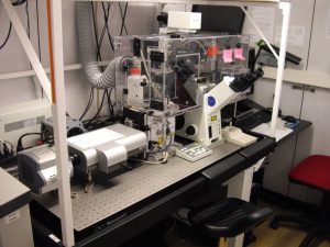
The platform Microscopy Imaging Côte d’Azur (« MICA ») (http://mica.unice.fr/) has a wide variety of state-of-the art microscopy equipment and expertise, accessible to all (internal and external) academic and industrial users. Our facility provides photonic, electron and atomic force microscopy, digital imaging (acquisition, treatment and analysis), flow cytometry, professional training and consultation.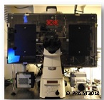 The two major foci of the platform are fluorescence and electron microscopy. A range of solutions exist at the MICA platform for studying different cell types and organisms at different scales. This platform accompanies the researcher in all aspects of microscopy, from training to equipment choice, experimental design, data acquisition and image/data analyses. Our goal is to assist users and in particular students and postdoctoral fellows with their research needs for qualitative and quantitative microscopy applications. We also assist with the production of publication-quality images and offer consultation on experimental approaches for use with our equipment. Furthermore, the platform engineers invest substantial time into methods development both with respect to equipment and image analyses. The equipment and the number of scientists, quality controllers, engineers and technicians dedicated to the optimal functioning of the MICA platform can be thus summarized:
- 19 specialized engineers and technicians
- 11 confocal microscopes (CLSM type) including 1 STED microscope
- 3 spinning disk confocal microscopes
- 3 electron microscopes (scanning and transmission)
- 16 video-microscopes including two TIRF/PALM microscopes
- 1 PALM micro-dissector
- 2 Nonlinear optical microscopes
- 1 Multi-beam multi-photon TrimScope
- 2 Atomic Force Microscopes, one which is coupled to an epifluorescence microscope
- 1 light-sheet ultramacroscopy system and 1 open-spim
- 9 FACs (6 analyzers and 3 cell sorters)
- 15 high-powered computers used for quantitative image analyses and treatments equipped with a range of software e.g. Metamorph, Amira, Imaris, Huygens, Volocity, Image J, Matlab etc.
The MICA platform regroups the imaging facilities of seven academic partners:
 Partner 1: The IPMC microscopy and flow cytometry facility provide a large panel of tools and methods, including all classical fluorescence microscopy methods (confocal, wide-field video-microscopy, homemade light-sheet ultramicroscope). Numerous teams study fast cellular phenomena in living samples and subcellular architecture. For this purpose, we use and develop specific tools to analyze the diffusion and interaction between molecules (FRAP, FCS and FCCS) as well as high-speed video-microscopy and image analyses. An increasing number of teams are doing dendritic spine morphology analysis thanks to a pipeline of confocal acquisition to a 3D analysis with the dedicated Imaris tool.
 Partner 2: The iBV communal microscopy platform have a strong investment in live cell fluorescence imaging at high-resolution coupled with high-speed image acquisition using both confocal and wide field microscopy systems. The development of new microscopy methods and equipment as well as tailor-made programs for quantitative image analysis is a major focus of this facility. We have developed a multimodal microscopy system that enables a substantial increase in resolution, i.e. beyond limits of diffraction of classical wide-field or confocal microscopes ultimately with two or more colors, which is available to users. Additional development of SHG microscopy and its applications is ongoing at the iBV.
 Partner 3: The C3M microscopy facility has 6 microscopes, providing a wide range of complementary techniques. It provides expert technical assistance (experimental design, acquisition and data analysis) to support investigators for basic and clinical research. In 2012, the platform has been certified ISO 9001:2008. The certification is to be renewed in 2016. In addition to confocal and wide field video microscopes, this facility has specific equipment such as TCSPC-FLIM, sreening and acquisition automation on a confocal system, a two-color TIRF microscope and a laser microdissector. The research teams at C3M work at the interface between fundamental and clinical research using samples ranging from cell culture to patient biopsies. The tissue imaging capabilities at C3M have been strengthened with the new histology facility, which is associated with the microscopy facility.
 Partner 4: The imaging platform of the LDBV is housed in the oceanographic observatory at Villefranche sur Mer. It is focused on understanding the embryonic development of key marine model organisms, e.g. jellyfish, sea urchins and ascidians, and have been pioneers in developing imaging techniques adapted for these species. They are currently employing the following imaging techniques: high speed confocal microscopy, wide field 4D fluorescence imaging, deconvolution, ratio-metric calcium imaging as well as image analysis, computer graphics & multimedia. They are members of the EMBRC consortium to share and supply expertise in that field. This facility is actually merging its activity with the other UMR of Oceanology in Villefranche sur Mer in order to share their equipments and knowledge in image analysis applied to automated in situ imaging of plankton.
 Partner 5: The CCMA is the only academic facility in Côte d’Azur dedicated to electron microscopy analyses. It is open to all laboratories of the University of Nice as well as all external academic institutions and private companies. Our long term expertise and newly acquired equipment allow us to provide several methods for analyzing cellular structures at high resolution from diverse biological samples (tissues and cells). Transmission Electronic Microscopy (TEM) and Scanning Electron Microscopy (SEM) are used daily. We are specialized in: 1) preparation of samples, including ultramicrotomy and cryo-ultramicrotomy, immunolabelling with gold particles, and freeze-fracture approaches; 2) qualitative and quantitative chemical analyses by back-scattering electron observations and energy-dispersive x-ray spectroscopy (EDS); 3) image analysis (quantification) in collaboration with partner 1. We are currently collaborating with partners 1, 2, 3 and 4, with respect to correlative microscopy methods, and the development of 3D imaging techniques, including tomography and serial section SEM (S3EM).
 Partner 6: The IRCAN is a research center (created in 2012) located at the Tour Pasteur in Nice. It assembles competitive research and clinical teams focusing on state-of-the art cancer and aging research ranging from molecular to clinical studies. The Pasteur-IRCAN Cellular and Molecular Imaging facility provides access to epi-fluorescence microscopes allowing high-content analysis and screening, fast wide-field coupled to integrated deconvolution and extensive cytogenetic analysis with specialized software. The facility recently upgraded its DeltaVision microscope with TIRF, FRET and high-resolution modules. IRCAN is also equipped with two AFM apparatuses, one of which is coupled to an epi-fluorescence microscope.
 Partner 7: The ISA of the INRA-PACA is comprised of 11 research teams mainly focusing on Plant/Insect-Microorganism interactions (symbiotic and pathogenic). Advanced microscopy dependent cell biology approaches are crucial for these studies involving the characterization of molecular and cellular processes responsible for the establishment, maintenance and rupture of these highly complex biotic interactions. The microscopy facility at ISA, created in 2004, and has built up extensive expertise in plant confocal imaging and cell biology particularly in the fields of plant-nematodes and microbes interactions.
Update-January, 2018


